


INTRODUCTION
Superior vena cava (SVC) syndrome is usually the result of the direct obstruction of the SVC by malignancies such as non-Hodgkin’s lymphoma, lung carcinoma. Moreover, SVC syndrome can be caused by infection such as syphilis and tuberculosis or by thrombosis in the vein itself. Recently the role of SVC thrombosis secondary to indwelling central venous device has drawn much attention. As breast cancer patients who receive chemotherapy increase, as the prevalence of installing chemoport increases. Kim et al. [1] analyzed 442 patients and reported immediate and delayed complication of totally implantable access devices. In his report, eleven patients experienced delayed complication such as, SVC thrombosis, catheter obstruction, pocket infection, etc. Treatment of SVC syndrome depends on the etiology of obstructive process. Thrombotic occlusion can be treated with appropriate pharmacologic agents. We describe the case of a breast cancer patient with SVC syndrome related with a central venous port catheterization for chemotherapy and treated with anticoagulation therapy only.
CASE REPORT
A 60-year-old female, afflicted with breast cancer, appealed recently aggravated face and neck swelling, fortunately she appealed neither dyspnea nor shortness of breath. Except tachycardia, physical examination showed no abnormal findings. Laboratory findings including complete blood cell count, electrolyte, coagulation profile were in normal limits.
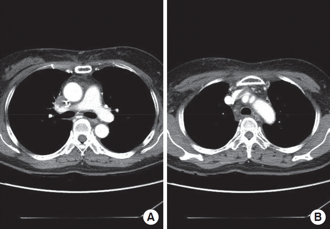
The patient underwent left breast-conserving surgery with sentinel lymph node biopsy followed by four cycles of AC (adriamycin 60 ㎎/㎡, cyclophosphamide 600 ㎎/㎡) chemotherapy and radiation therapy. After the first AC chemotherapy, central venous port was placed into the right subclavian vein. During radiation therapy she got regular port irrigation with heparin diluted fluid. Accomplishing radiation therapy, she was scheduled to get 18 cycles of trastuzumab therapy for a year. After the 14th trastuzumab therapy, she appealed chest tightness, face and neck swelling, so we conducted contrast-enhanced computed tomography (CT) of the chest and CT showed thrombotic filling defect of the SVC and proximal left brachiocephalic vein (Figure 1). Considering her clinical symptoms and CT findings, we applied low molecular weight heparin 60 mg every 12 hours to the patient for 10 days. Ten days later, we checked follow-up chest CT angiography.
Follow-up CT angiography showed increased amount of thromboembolism in SVC and left brachiocephalic vein and it showed more improved edematous soft tissue change, but her symptoms were not improved (Figure 2). So we considered fibrinolytic therapy, and we transferred the patient to the vascular department for fibrinolytic treatment.
Fourteen days later, before conducting fibrinolytic treatment, we rechecked upper extremity venous CT angiography (VCTA), it showed more decreased thrombotic filling defect at SVC, left brachiocephalic vein, and subclavian vein, at the same time, patient’s symptoms were more relieved. Instead of conducting fibrinolytic treatment, we continued low molecular weight heparin treatment combined with warfarin medication. One week later from last exam, we rechecked upper extremity VCTA and it showed no filling defect at any vessels mentioned before (Figure 3). We removed central venous port and the patient was discharged with anticoagulation (warfarin with a target international normalized ratio between 2.5 and 3).
Two weeks later, she visited outpatient clinic and we checked vascular flow using Doppler ultrasonography, it showed no disturbance of blood flow and her irritating symptoms disappeared.
DISCUSSION
Central venous port catheter is frequently conducted in the fields of oncology, parenteral nutrition, fluid and factor replacement, and frequent blood sampling. Moreover central venous port catheterization is commonly applied to breast cancer patients who need long-term chemotherapy and additional intravenous treatment. It is reported that the incidence of noninfectious complication such as obstruction of the distal extremity of the catheter and deep vein thrombosis, ranges from 7% to 50% [2]. The formation, growth, and dissolution of venous thrombosis are determined by several thrombogenic factors (duration of catheterization, physical properties of the catheter, nature of infused fluid). The common symptoms of SVC syndrome are facial swelling, dyspnea, coughing and arm swelling. In spite of high thrombotic incidence, symptomatic massive thrombosis causing SVC syndrome is not common. Although central venous port catheter was the main cause of catheter related thrombosis in our patient, the breast cancer itself must have contributed to the extent of thrombosis.
Diagnostic tools of SVC syndrome are composed of clinical suspicion and venography or CT angiography. Trials have reported the use of Doppler flow studies [3], echocardiography, and digital subtraction angiography [4]. At first, we conducted CT angiography, because it is easy and rapid not only for the diagnosis but also for comparison of treatment courses.
There are different alternatives for treatment of SVC syndrome. Most patients with SVC syndrome secondary to malignancy are treated nonoperatively, with radiotherapy, chemotherapy, or both. Patients who develop catheter-related SVC obstruction are candidates for anticoagulant treatment and may require catheter removal. The standard delivery of the drug is by infusion catheters that are centered in the thrombus. Thrombolysis has been successful in about 75% of patients when initiated within 2 days after the onset of symptoms, but it is rarely successful after 10 days [5]. Percutaneous endovascular stents have been done with success to relieve obstruction to blood flow, and these stents have become a surrogate option for treating patients with SVC syndrome. Operative therapy is rarely indicated. Surgery has a role in treating highly selected patients such as those with complete occlusion of the SVC, severe refractory symptoms, and thrombosis of venous collaterals [6]. Anticoagulation therapy and thrombectomy have been the mainstream of treatment to prevent thrombosis propagation and relive symptoms in patients with catheter related thrombosis [7]. Doty et al. [8] reported that a spiral vein bypass graft relieved SVC syndrome and provided excellent long-term patency in 16 patients. Guijarro Escribano et al. [9] reported a case of SVC syndrome associated with central venous catheter use for chemotherapy, which was successfully treated with catheter-directed urokinase infusion. Boza et al. [10] presented a case, successfully treated with thrombolysis, angioplasty, and stent placement. Fortunately, in our patient, she was successfully treated with anticoagulation therapy without invasive treatment.
In conclusion, central venous port catheterization could result in severe thrombosis which may be further complicated by combined cancers, emphasizing the need for close observation of patients having a central venous catheter. Anticoagulation therapy may be applied for complete resolution of thrombosis especially when diagnosed early. Surgeons also speculate the possibility of SVC syndrome in patients who have central venous port catheter.